8APEM, Kanazawa, 2004
DeConvHAADF: Software Cs-corrector for STEM-HAADF Microscopy
Kazuo Ishizuka
HREM Research Inc, Matsukazedai, Saitama 355-0055, Japan
ishizuka@hremresearch.com
Summary
The scanning transmission electron microscope (STEM)
is able to resolve atomic columns of crystalline materials by using a
high-angle annular dark-field (HAADF) detector. Since HAADF imaging is believed
to be incoherent and described as a convolution of a scattering distribution
and an electron probe, we can improve image resolution by deconvolution of the
STEM-HAADF image with an electron probe function. In this presentation we
report improvement of spatial resolution of STEM-HAADF images by deconvolution
procedures based on both maximum entropy routine and Richardson-Lucy routine.
It has been also demonstrated that pre-convolution with a Gaussian removes
quantum/granular noise and yields a more reliable result.
Keywords: STEM-HAADF, deconvolution, maximum entropy,
Richardson-Lucy, MEM
The scanning transmission electron microscope (STEM) is able to resolve atomic columns of crystalline materials by using a high-angle annular dark-field (HAADF) detector [1]. There has been effort to improve resolution further by correcting spherical aberration of the objective lens [2]. However, the HAADF imaging is believed to be incoherent and described as a convolution of a scattering distribution and an electron probe [3]. Therefore, we can improve image resolution by deconvolution of the STEM-HAADF image with an electron probe function. Cambridge maximum entropy program (MaxEnt) [4] was thus adapted and tested on STEM-HAADF images of simple structures [5]. In this case an object function is assumed to be made of delta functions, which is a good assumption for a stellar object. Another deconvolution technique recently developed in astronomy to correct a defect of Hubble space telescope images uses Richardson-Lucy algorithm [6]. In the field of electron microscopy, this technique was applied to EELS [7] and CBED [8]. We have developed deconvolution procedures to improve energy resolution of EELS (DeConvEELS) [9]. There, we implement both maximum entropy (ME) routine based on Collin's approach [10] as well as Richardson-Lucy (RL) routine. In this presentation we report improvement of spatial resolution of STEM-HAADF images by deconvolution procedures based on the same principles.
Both ME and RL algorithms are based on Bayes' theorem of probability to reconstruct the most probable image, which is consistent with the observed data. The procedures estimate a predicted image by convoluting it with the probe function. Since they do not perform a direct deconvolution, they are not so much affected by noise compared with the Fourier deconvolution. It is interesting to note that these deconvolution routine can eliminate also an artificial convolution applied to reduce the noise in the image, which is not possible by using the Fourier deconvolution. Since both procedures iteratively estimate the most probable image, important issues are how to control their convergence and when we stop the processing. There are a few parameters for these procedures that may be optimized for each image to be processed. However, it may be worthwhile to note that the following results were obtained by using default parameters.
Figure 1 shows a quasi-crystal using JEOL 2010F [11] (200 kV, Cs: 0.5 mm, probe forming angle: ~12 mrad, ADF detector: 50 - ~110 mrad.). Result obtained by the ME and RL deconvolutions using a calculated probe at the Scherzer conditions are shown in Figure 2. The resulting images show very substantial reduction in noise. Here, the original data was convoluted with a Gaussian before deconvolution (pre-convolution).
We have developed stable and easy-to-use deconvolution procedures for STEM-HAADF images (DeConvHAADF) [12]. It has been demonstrated that pre-convolution with a Gaussian removes quantum/granular noise and yields a more reliable result.
References Figure 1. Original quasi-crystal
image obtained using JEOL 2010F (200 kV, Figure 2. ME (left) and RL (right) deconvolutions with
pre-convolution with a Gauusian
[1] S.J. Pennycook, D.E. Jesson, Phys. Rev. Lett. 64 (1990) 938.
[2] O.L. Krivanek, N. Dellby, A.R. Lupini, Ultramicroscopy 78 (1999), 1-11
[3] S.J. Pennycook, D.E. Jesson, Ultramicroscopy 37 (1991) 14-38.
[4] S.F. Gull, J Skilling, IEEE Proc131F (1994) 646-659.
[5] S.J. Pennycook et al., Proc. 10th Pfefferkorn
Conf., Cambridge, 1992, 233-243; H.S. von Harrach, D. Krause, Proc. 13th
Intern. Congress on Electron Microscopy (1994), Paris, Vol. 1, 479-480.
[6] W.H. Richardson, J Opt Soc Am 62 (1972) 55;
L.B. Lucy, Astrophysical Journal 79 (1974) 745.
[7] A. Gloter et al., Ultramicroscopy .96 (2003) 385-400.
[8] J.M. Zhuo, Microscopy Research and Technique 49 (2000) 245.
[9] K. Ishizuka, K. Kimoto, Y. Bando, Microsc
Microanal 9 (Suppl 2) (2003) 832-833; DeConvEELS is commercially available from
HREM Research Inc (www.hremresearch.com)
[10] D.M. Colin, nature 298 (1982) 49-51.
[11] E. Abe, S.J. Pennycook, A.P. Tsai, Nature 421 (2003) 347-350.
[12] DeConvHAADF is commercially available from HREM Research Inc (www.hremresearch.com).
[13] The author greatly acknowledges Dr. E. Abe for
providing the quasi-crystal data to test the procedures developed here.
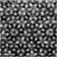
Cs: 0.5 mm, probe forming angle: ~12 mrad, ADF detector: 50-~110
mrad.)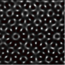
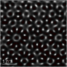
(10 pixels). It is clear that both
deconvolution procedures yield almost identical results.