(WinHREMTM/MacHREMTM)
|
High Resolution Electron Microscope Image Simulation Programs |
|||||||||||||||||||||||||||||||||
 |
|||||||||||||||||||||||||||||||||
|
High Resolution Electron Microscope Image Simulation Programs |
|||||||||||||||||||||||||||||||||
High Resolution Electron Microscopy (HREM) becomes an indispensable tool for understanding material properties or evaluating new materials at the level of atomic resolution. Due to increased demands for research and development of the new materials, an image simulation acquires more importance than ever before. WinHREMTM/MacHREMTMis a suite of the high-resolution electron microscope image simulation programs that will run on Windows PC or PowerMacintosh. |
|||||||||||||||||||||||||||||||||
| User Friendly Graphical Interface | |||||||||||||||||||||||||||||||||
|
WinHREMTM/MacHREMTMemploys user friendly Data Generation Utilities based on the Graphical User Interface for Windows or Mac OS.
WinHREMTM/MacHREMTM is general-purpose software that can be used to simulate all the images expected from any crystal systems, defect structures and interfaces. Although data generation for such general-purpose software normally becomes complex, a novice user can easily generate his/her data by using the graphical Data Generation Utilities with minimum requirements for the special knowledge.
|
|||||||||||||||||||||||||||||||||
| Reliable and Efficient Algorithm | |||||||||||||||||||||||||||||||||
|
Since electron microscope images critically depend on an electron-specimen interaction as well as aberrations of image forming lenses, the treatment of scattering based on dynamical theory and the treatment of aberration based on wave-optical theory are mandatory.
WinHREMTM/MacHREMTM emerges from the HREM image simulation programs based on FFT multislice technique developed at Arizona State University, USA (see References). This is one of the most reliable and efficient HREM image simulation programs.
Features of WinHREMTM/MacHREMTM
K. Ishizuka and N. Uyeda, A New Theoretical and Practical Approach to the Multislice Method, Acta Cryst. A33 (1977) 740; K. Ishizuka, Contrast Transfer of Crystal Images in TEM, Ultramicroscopy 5 (1980) 55; K. Ishizuka: A practical approach for STEM image simulation based on the FFT multislice method, Ultramicroscopy 90 (2001) 71-83; K. Ishizuka, Multislice Formula for Inclined Illumination, Acta Cryst. A38 (1982) 773-779. |
|||||||||||||||||||||||||||||||||
| High Quality Image Output | |||||||||||||||||||||||||||||||||
|
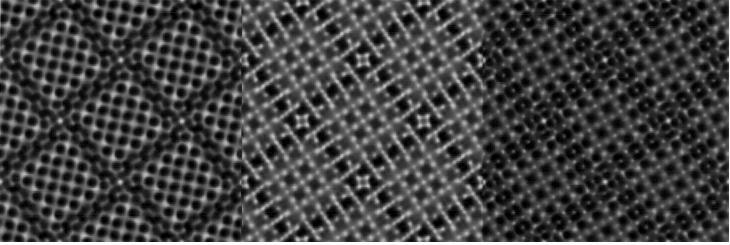
Numerical data such as projected potential, wave function propagating the specimen, simulated image intensities could be converted into a standard image format for Windows or Mac OS (Bit map or PICT) and printed as high quality pictures by using Output Graphic Utilities. Photographic quality images as shown below could be obtained by using a high-quality printer. |
|||||||||||||||||||||||||||||||||
|
|||||||||||||||||||||||||||||||||
|
Simulated HREM images for tungsten niobate at 200 kV with Cs = 0.5 mm Assumed thickness is 3.8nm; Defocuses from left to right are 42nm, 65nm and 83nm (under-focus) |
|||||||||||||||||||||||||||||||||
| Powered by Optional Functions | |||||||||||||||||||||||||||||||||
|
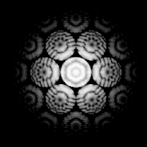
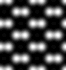
WinHREMTM/MacHREMTM could be extended its capabilities to simulate convergent-beam electron diffraction (CBED) patterns, diffuse scattering intensity distributions as well as scanning transmission electron microscope (STEM) images including high-resolution high-angle annular dark-field (HAADF) images by adding corresponding optional functions. |
|||||||||||||||||||||||||||||||||
|
|
||||||||||||||||||||||||||||||||
|
Simulated CBED pattern for Si [111] |
Simulated HAADF image for GaAs [011] |
||||||||||||||||||||||||||||||||
| WinHREMTM/MacHREMTM Specifications | |||||||||||||||||||||||||||||||||
|
|||||||||||||||||||||||||||||||||
|
|||||||||||||||||||||||||||||||||
| Please Contact to: | |||||||||||||||||||||||||||||||||
 Send your inquiries about the price for a new purchase, an upgrade or a cross-upgrade to: support@hremresearch.com |
HREM Research Inc. 14-48 Matsukazedai Higashimatsuyama 355-0055, JAPAN TEL/FAX 0493-35-3919 web site: www.hremresearch.com email: support@hremresearch.com |
||||||||||||||||||||||||||||||||